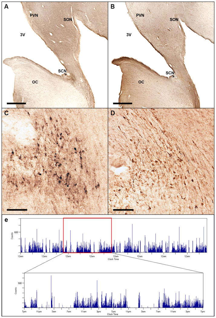
Figure 1.
Representative anatomic and actigraphic data. Immunohistochemical staining of human suprachiasmatic nucleus for vasoactive intestinal peptide (VIP) (A, C) and arginine vasopressin (AVP) (B, D). Low power (a, b, scale bar = 1 mm) and high power views (c, d, scale bar = 100 μm) from a single subject in consecutive sections. (E) Visualization of actigraphy data from an illustrative participant. (Inset: detail view of 48 hours of this recording). OC = Optic Chiasm; PVN = Paraventricular Nucleus; SCN = Suprachiasmatic Nucleus; SON = Supraoptic Nucleus; 3V = 3rd ventricle.

