Fig. 4.
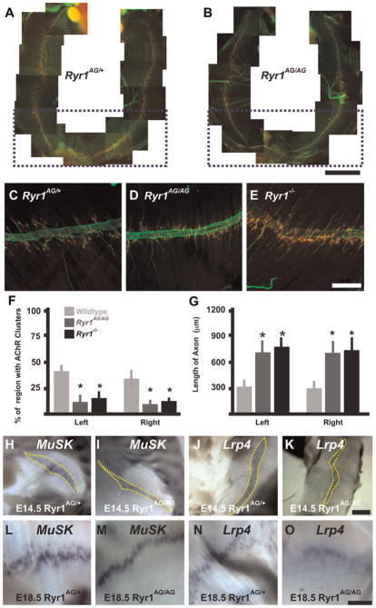
Narrowed AChR cluster distribution and increased motor axon outgrowth in Ryr1AG/AGdiaphragms. (A, B) E18.5 whole diaphragms show clustered AChRs that form neuromuscular junctions visualized with α–bungarotoxin (red) and axons with synaptophysin and neurofilament (green; scale bar in A, B=1 mm). (C–E) Left ventral quadrants of diaphragms (scale bar=200 μm). (C) In Ryr1AG/+ diaphragms, AChR clusters extend along the axon branches. (D, E) In Ryr1AG/+ and Ryr1−/− diaphragms, AChR clusters surround the fasciculated nerve but do not extend along the axon branch. (F–G) Quantification in the ventral quadrant (outlined by purple boxes in A, B) of AChR cluster regionalization (F) and axon length (G) (n=5 for each). H–O, At 14.5 (H–K, scale bar=100 μm) and at E18.5 (L–O, scale bar=200 μm), MuSK and Lrp4 (yellow dashes) are expressed in the central region of Ryr1AG/+ and Ryr1AG/AG diaphragms.

