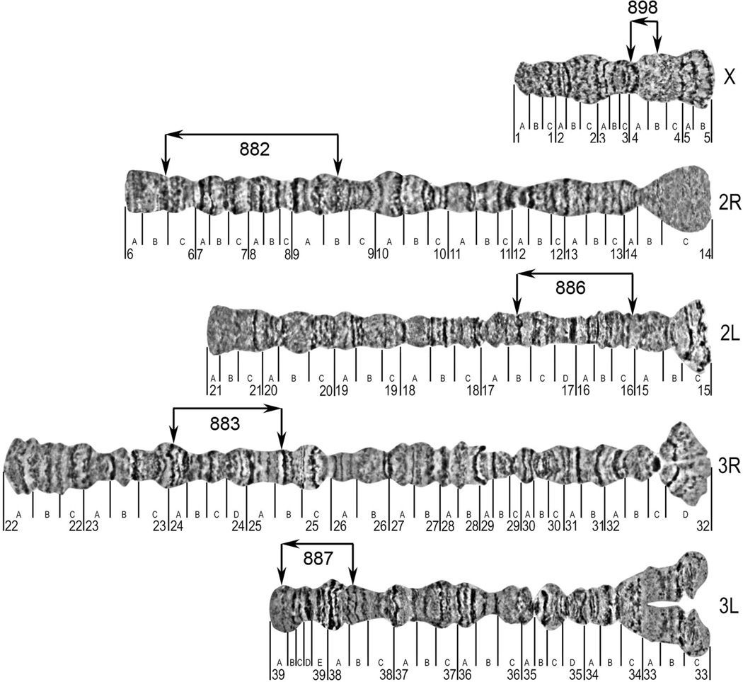
Figure 2. A standard cytogenetic photomap of ovarian nurse cell chromosomes of An. atroparvus.
The lines below the chromosomes indicate the boundaries of numbered and lettered divisions and subdivisions respectively. The locations of DNA probes from the start and the end of the genomic supercontigs are indicated by vertical arrows above the chromosomes. The orientations of the genomic supercontigs are shown by horizontal arrows above the chromosomes.

