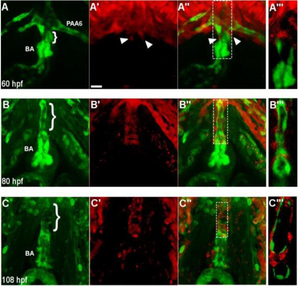
Fig. 4.

Neural crest-derived cells surround the endothelium of the ventral aorta and bulbus arteriosus. (A-A’’) A confocal projection of a ventral view of the BA and VA of a NC:NfsB-mCherry; kdrl:GFP embryo at 60 hpf (anterior toward top). Arrowheads point to neural crest cells migrating onto the VA from the 6th aortic arch artery. (B-C’’) Confocal projections of BA and VA of a NC:mCherry; kdrl:GFP embryo at 80 hpf (B-B’’’) and 108 hpf (C-C’’’) show the progression of mCherry+ cells migrating along the VA. A’’’, B’’’ and C’’’, Single z section of area boxed in A”, B’’ and C”, respectively. Brackets mark the ventral aorta (VA); BA, bulbus arteriosis; PAA6, pharyngeal arch artery 6, scale bar = 20µm.
