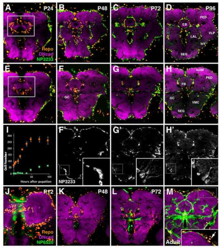
Figure 8.
Pattern of secondary neuropil glia throughout metamorphosis. (A–H) Z-projections of frontal confocal sections of pupal brains in which sALG are labeled by NP3233-Gal4 (green); the neuropil is visualized by anti-DN-cadherin (magenta), and glial cell nuclei are labeled by anti-Repo (orange). sALG from (F–H) are shown in grayscale with high magnification insets of boxed regions in (F’-H’). Panels of first row (A–D) show z-projections at level of ellipsoid body (EB); panels of second and third row (E–H, F’-H’) show slightly more posterior level at fan-shaped body (FB). Boxed regions in (A, E) depict the domains in which cell counts were conducted (I); (I) total Repo-positive cells associated with the central complex (i.e. EB, FB, noduli) are shown in orange, fraction of Repo-positive cells which co-express NP3233-Gal4 is shown in light green (n>3 for each time point). At early and mid-pupal stages, sALG spread out around neuropil surface and central complex primordium while at the same time increasing in number (A, E, B, F–F’). sALG processes do not yet enter the neuropil at significant numbers (see inset in F’). During the second half of the pupal stage (C, G–G’), radial processes emanate from the sALG into the neuropil (see inset in G’). Glial processes form a rich network of branches during the last day of metamorphosis (D, H–H’; see inset in H’). (J–M) Developmental time course of primary and secondary ensheathing glia (sEG) from P12 to adult at the level of the fan-shaped body (FB) and great commissure (GC). EG are labeled by NP6520-Gal4 in green, neuropil is labeled by anti-DN-cadherin in magenta, and glial cell nuclei are labeled by anti-Repo in orange. At P12 (J) primary EG of the larva are still present. During mid-metamorphosis (K), NP6520-positive pEG are no longer seen. NP6520 expression in secondary EG appears first around the central complex (L), followed by expression at the neuropil surface (M). Clusters of NP6520-Gal4 negative neuropil glia, interpreted as sALG, occupy regions of low sEG density at the adult neuropil surface (M, inset; arrow). Anatomical abbreviations found in Table 1.
Bar: 50µm

