
FIGURE 1.
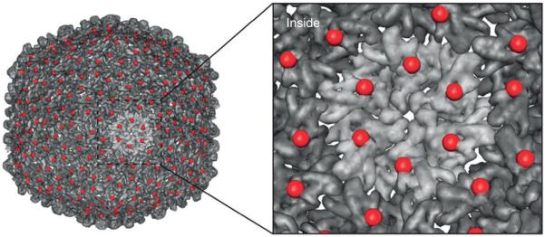
The symmetry of VLPs reflects a single change over the entire particle. The VLP from bacteriophage P22 is shown with a serine to cysteine point mutation at amino acid position 39, displayed as a red sphere. This single mutation provides a new site at each of the 420 coat proteins that make up the capsid. (Reprinted with permission from Ref 4. Copyright 2012 Nature Publishing Group)