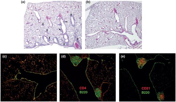
FIGURE 4.
iBALT readily forms in the murine lung after i.n. administration of sHSP. (a) iBALT structures,indicated by arrows, emerge adjacent to airways and blood vessels after five administrations of sHSP compared to a PBS control (b). Stained fluorescence microscopy reveals that, compared to a control (c), iBALT structures contain (d) CD4+ T-cells, B220+ B cells, and (e) CD21+ follicular DC. (Reprinted with permission from Ref 43. Copyright 2009 Public Library of Science (PLoS))

