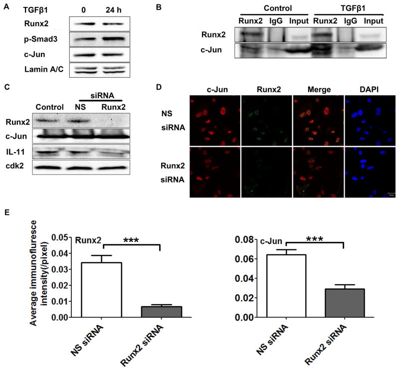
Fig. 5. Runx2- and TGFβ1-induced c-Jun form a complex in the nucleus.
(A) Western blot showing expression of Runx2, p-Smad3, and c-Jun in PC3-H cells at the indicated times in the presence or absence of TGFβ1 treatment. Lamin A/C was used as internal control. (B) Runx2 co-immunoprecipitation in PC3-H cells after TGFβ1 treatment. PC3-H cells were treated with TGFβ1 or DMSO-control, then immunoprecipitated using Runx2 or IgG control. The Western blot shown was probed with Runx2 and c-Jun antibodies, as indicated. (C–E) Runx2 silencing in PC3-H cells. (C) Western blot analyses of Runx2, c-Jun, IL-11 protein in non-transfected (control) cells or 48 h after transfection with Runx2 siRNA or a non-silencing (NS) control siRNA. Cdk2 was used as loading control. (D) Representative confocal images 48 h after delivery of Runx2 or a non-silencing (NS) control siRNA (Scale Bar = 20 μm). (E) Quantification of the immunofluorescence signal in panel D calculated using the ImageJ software package. Data are mean ±s.d, calculated using the average intensity of fewer random fields of view (~10 cells/field) in three independent experiments. Statistical analysis (T-test with Welch’s correction) was used to compare the values of cells transfected with NS siRNA or Runx2 siRNA: ***p<0.001.

