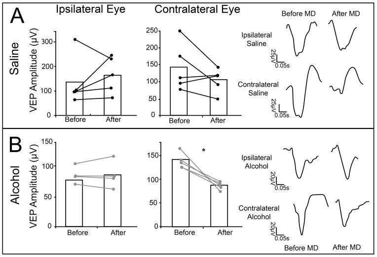
Figure 6.
Changes in individual eye responses after 3 days of MD. After 3 days of MD, saline treated animals exhibited no change in response amplitude of the ipsilateral eye (133.73μV ±44.35; 163.75μV ±33.71; n = 5), yet the contralateral eye (169.51μV ±44.3; 146μV ±22.6; n = 5) exhibited a trend toward decreased response amplitude (A). This change can be seen in VEP responses from a representative animal. Ethanol exposed animals also showed a significant decrease in their contralateral eye responses (126.5μV ±7.56 to 90.7μV ±4.44, n=4) after MD, but no changes were seen in ipisilateral responses (77.43μV ±6.09; 5.01μV ±10.09; n= 5) (B). This change can be seen in VEP traces from a representative animal. * p = 0.01.

