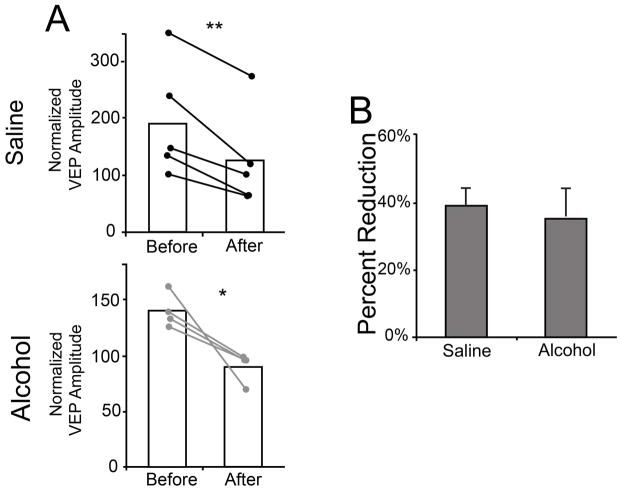
Figure 7.
Normalized contralateral eye responses after 3 days of MD. When contralateral eye VEPs are normalized to ipsilateral eye responses, the decrease in contralateral eye responses for all groups becomes apparent (A). With this data transformation, saline and alcohol treated animals exhibit a similar percent change in VEP amplitude from before to after MD (B). * p = 0.04, ** p = 0.007

