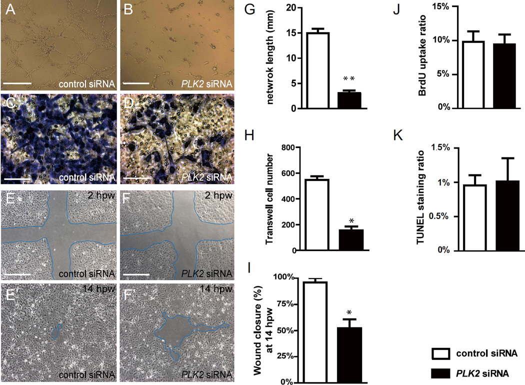
Figure 3. PLK2 regulates endothelial cell migration.
(A, B) EC tube formation assays, (C, D) transwell EC migration assays, and (E–F’) wound healing EC assays show that when compared to control siRNA knockdown, PLK2 siRNA knockdown in HUVECs results in (B and G) decreased EC network/tube formation (p = 0.0028), (D and H) reduced EC migration formation (p = 0.0185), and (F, F’ and I) slower EC wound healing/closure, respectively (p = 0.0114). (J–K) Quantitative measurements of the percentage of (J) BrdU positive cells and (K) TUNEL staining positive cells in control and PLK2 siRNA transfected HUVECs show that PLK2 knockdown does not affect HUVEC cell proliferation (p = 0.6007) and apoptosis (p = 0.7970). (A, B, E–F’) Scale bar, 1.6 mm. (C, D) Scale bar, 400 µm. hpw – hours post wounding. Mean +/− s.e.m. *p<0.05, **p<0.01 by Student's t-test.

