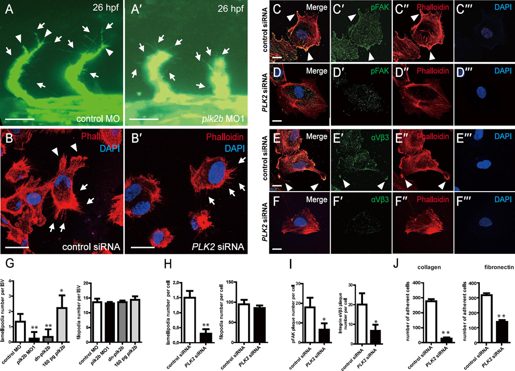
Figure 4. PLK2 regulates endothelial cell lamellipodia and cell adhesion formation.
(A-A’) 26 hpf plk2b MO1 injected Tg(fli1a:eGFP) fish (n = 31/40) exhibit fewer lamellipodia, and extending intersomitic vessels compared to age-matched control MO injected Tg(fli1a:eGFP) fish (n = 0/48). Scale bar, 40 µm. Top, dorsal longitudinal anastomotic vessel; bottom, dorsal aorta/cardinal vein. (B-B’) Phalloidin staining reveals that PLK2 siRNA transfected HUVECs display reduced lamellipodia when compared to control siRNA transfected HUVECs. Scale bar, 20 µm. Immunostaining of (C-C”’, D-D”’) pFAK and (E-E”’, F-F”’) integrin αVβ3 shows that focal adhesions and integrins, respectively, are localized to the lamellipodia of migrating/extending (C, E) control siRNA transfected HUVECs, but they fail to organize and aggregate in (D, F) PLK2 siRNA transfected HUVECs. Scale bar, 10 µm. (G) Quantitative measurements of the number of lamellipodia and filopodia reveal that zebrafish plk2b knockdowns have reduced lamellipodia but relatively the same number of filopodia (finger-like protrusions crossing the cell edge) when compared to controls. Conversely, zebrafish RNA injection of 160 pg plk2b resulted in more lamellipodia only. (H) Quantitative measurements of the number of lamellipodia and filopodia reveal that human PLK2 knockdowns have reduced lamellipodia (p = 0.0023) but relatively the same number of filopodia (p = 0.2615) when compared to controls. (I) Quantitative measurements of the number of pFAK (p = 0.0143) and integrin αVβ3 plaques (p = 0.0224) reveals that knockdown of PLK2 reduced the number of cell adhesions in HUVECs. (J) EC adhesion assays show that PLK2 siRNA HUVECs adhered less to type I collagen (p = 0.0017) and fibronectin-coated coverslips (p = 0.0061) compared to control siRNA HUVECs. Arrowheads and arrows point to lamellipodia and filopodia, respectively. Red – phalloidin/actin staining; Blue – DAPI staining; Green – (A) GFP, (C–D) pFAK, or (E, F) integrin αVβ3. Mean +/− s.e.m. *p<0.05, **p<0.01 by ANOVA for G and Student's t-test for H–J.

