Fig. 3.

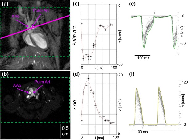
In vivo validation of 4D PC FLASH MRI in mice. a, b Anatomical overview obtained with self-gated FLASH showing images of the mouse heart in coronal (a) and axial view (b), wherein the solid purple line in (a) corresponds to the selected slice in (b) in the slice-selective 2D PC MRI experiments. For 4D PC MRI experiments, a three-dimensional FOV covering the whole heart and great vessels was chosen (green dashed lines). b illustrates the locations in which the velocities are calculated in the ascending aorta (AAo) and the pulmonary artery (Pulm Art). c–f present the temporal evolution of peak flow velocities in velocity–time curves for the two vessels [Pulm Art (c, e) and AAo (d, f)] for 4D PC MRI (c, d) and ultrasound (e, f). The green and yellow lines in e and f are the envelopes of the ultrasound velocity data.
