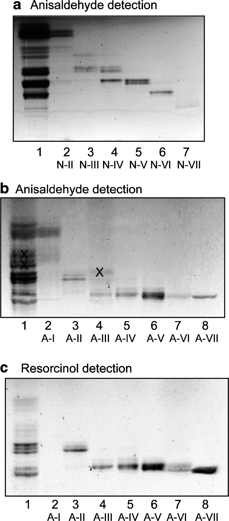
Fig. 2.
Separation of glycosphingolipids from moose I small intestine. Thin-layer chromatogram of non-acid (a) and acid (b and c) glycosphingolipid fractions isolated from moose I small intestine. The chromatograms were eluted with chloroform/methanol/water 60:35:8 (by volume), and detection of glycosphingolipids was done with anisaldehyde (a and b), and the resorcinol reagent (c). The lanes on (a) were: Lane 1, total non-acid glycosphingolipids of moose I small intestine, 40 μg; Lanes 2–7, fractions N-II – N-VII from moose I small intestine, 4 μg/lane. The lanes on (b and c) were: Lane 1, total acid glycosphingolipids of moose I small intestine, 40 μg; Lanes 2–8, fraction A-I – A-VII from moose I small intestine, 4 μg/lane. The bands marked with X on (b) are non-glycosphingolipid contaminants

