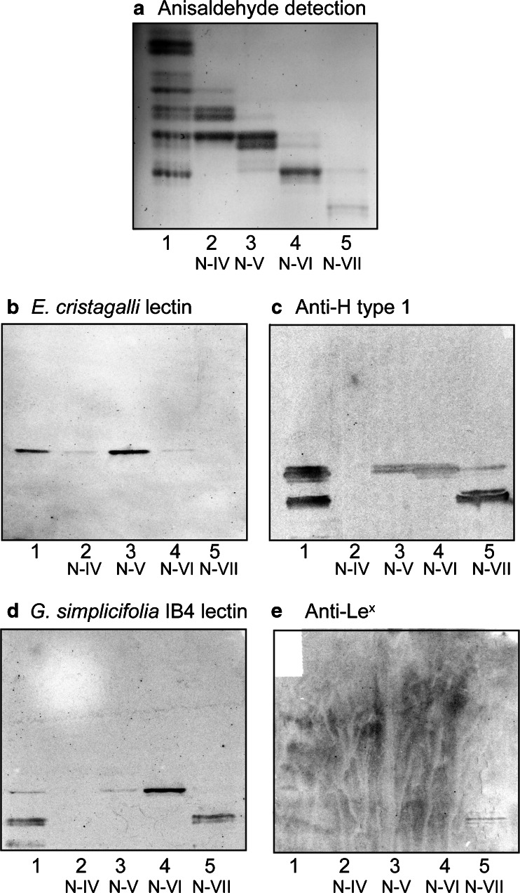
Fig. 4.
Binding of lectins and monoclonal antibodies to the non-acid glycosphingolipid subfractions isolated from moose I small intestine. Thin-layer chromatogram after detection with anisaldehyde (a), and autoradiograms obtained by binding of E. cristagalli lectin (b), the monoclonal anti-H type 1 antibody 17–206 (c), G. simplicifolia IB4 lectin (d), and the monoclonal anti-Lewisx antibody P12 (e). The glycosphingolipids were separated on aluminum-backed silica gel plates, using chloroform/methanol/water 60:35:8 (by volume) as solvent system, and the binding assays were performed as described under “ Materials and methods”. Autoradiography was for 12 h. The lanes were: Lane 1, total non-acid glycosphingolipids of moose I small intestine, 40 μg; Lanes 2–5, fraction N-IV – N-VII from moose I small intestine, 4 μg/lane

