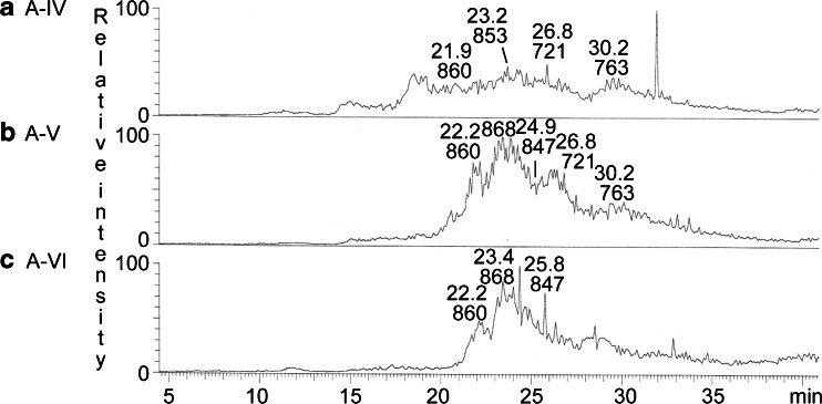
Fig. 9.
LC-ESI/MS of the acid glycosphingolipids from moose I small intestine. a Base peak chromatogram from LC-ESI/MS of fraction A-IV from moose I small intestine. b Base peak chromatogram from LC-ESI/MS of fraction A-V from moose I small intestine. c Base peak chromatogram from LC-ESI/MS of fraction A-VI from moose I small intestine

