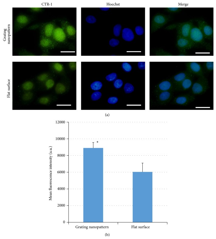
Figure 6.
(a) Immunofluorescent staining of copper transporter-1 (green excitation λ = 448 nm) in HepG2 cells cultured on grating nanopattern with total dimensions (240 nm) and grating shapes (upper panel) and flat surface (lower panel) (scale bar = 20 μm). (b) Calculated mean fluorescence intensity that shows a significant increase in the fluorescence intensity for cells cultured over grating nanopattern substrate compared to flat surface. ∗Statistically significant at (P < 0.05).

