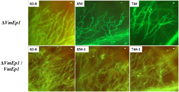
FIGURE 7.

Epifluorescence microscopy of tissue colonization during infection. Boundary-zones (∼0.5 cm2) of inoculated leaves were sampled at 60 hpi. All treatments were performed with at least five replicates, and all experiments were repeated three times. Bar = 10 μm.
