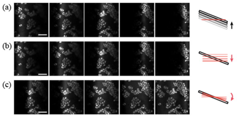
Fig. 2.

Full-field visualization of a slide of HeLa cells oriented at a 15° inclination. Z-stack images are acquired (a) by translating the sample in 20 μm steps along the optic axis and (b) by moving the z-scanning mirror. (c) The dynamic motion of the z-scanning mirror is synchronized with the fast axis of lateral scan and the tilt of image plane is gradually adjusted by increasing the amplitude. Scale bar, 100μm.
