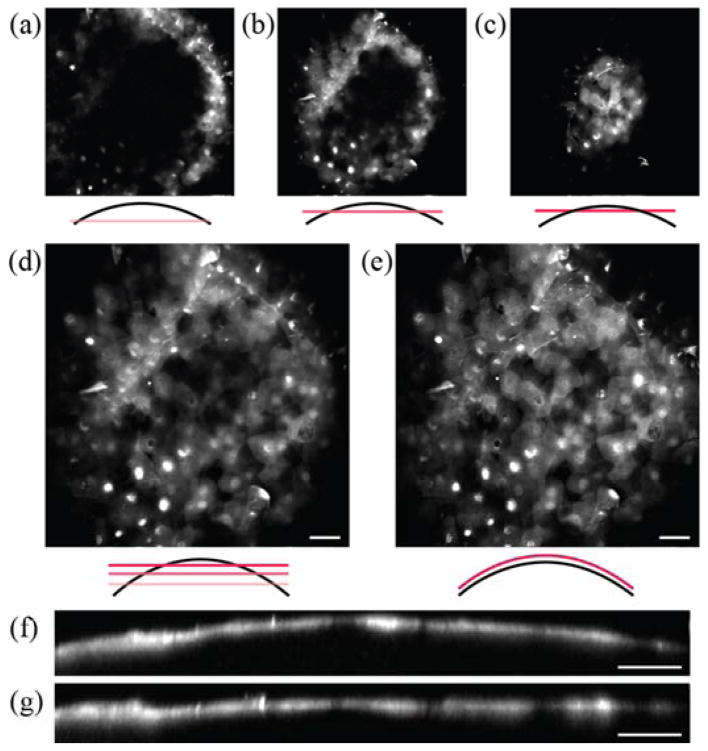
Fig. 4.
The cornea of mouse eye imaged by adaptive field microscopy. The superficial epithelium is visualized. (a), (b), and (c), are z-sections that are 10 μm apart in depth with field shaping OFF. (d) The average z-projection image over 20-μm depth compared with (e) a single z-section with field shaping ON. (f) and (g) are the axial view of the corneal epithelium with and without field shaping. Scale bars, 40μm.

