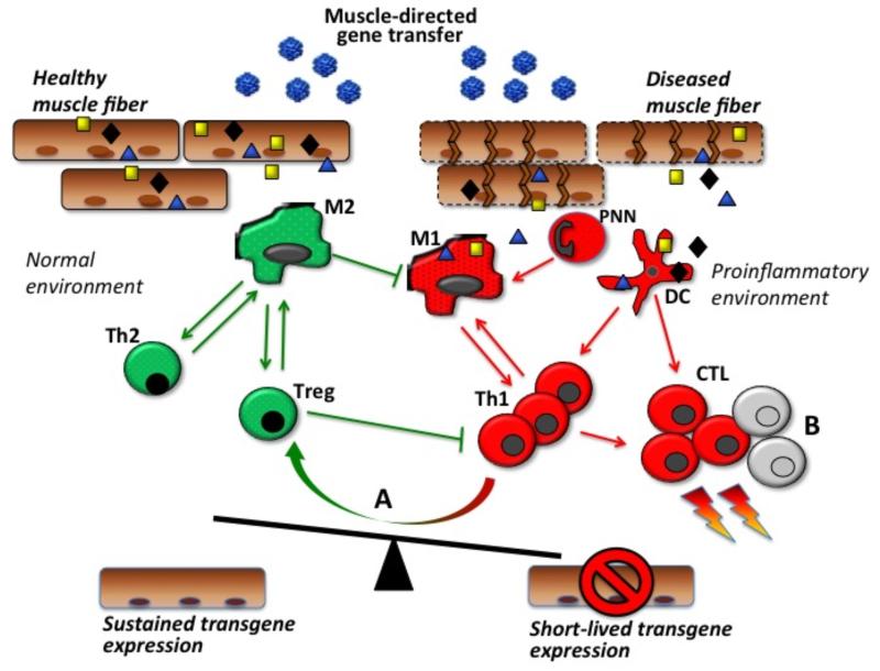
Fig. (2). Transgene expression in muscle and the balance between tolerance and immunity.
The release of immunogenic antigens by leaky, damaged muscle fibers upon rAAV intramuscular administration and their uptake by dendritic cells (DC) contribute to the activation of immune responses and destruction of transduced muscle fibers. The proinflammatory conditions provided by the diseased muscle environment drive the activation of effector cells including type 1 macrophages (M1), granulocytes (PNN), type 1 CD4+ T helper cells (Th1), and CD8+ cytotoxic T lymphocytes (CTL). In a normal muscle environment, type 2 macrophages (M2), type 2 CD4+ T helper cells (Th2), and regulatory T cells (Treg) may dampen the effector immune response and contribute to sustained transgene expression. (A) in inflamed muscle, resident Tregs may be induced and contribute to modulate immune responses at the muscle level; similarly (B) effector T cell exhaustion and/or apoptosis may contribute to limit muscle inflammation.

