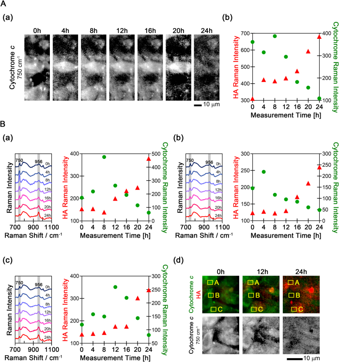
Figure 3. The temporal changes of cytochrome c and HA Raman signals from osteoblasts.
(A) The temporal changes in cytochrome c are shown as (a) Raman images and (b) Raman intensity plots. The Raman intensity of HA is also shown in (b). (B) The temporal changes in cytochrome c and HA Raman intensity in three additional locations Panels (a–c) indicate the temporal changes in the averaged Raman spectra and Raman intensity of cytochrome c and HA in the three cells. The averaged Raman spectra were obtained from the regions boxed in yellow labeled A, B, and C in (d). Panel (d) shows the time-lapse merged Raman images of cytochrome c (750 cm−1, shown in green) and HA (956 cm−1, shown in red) and the individual Raman images of cytochrome c.

