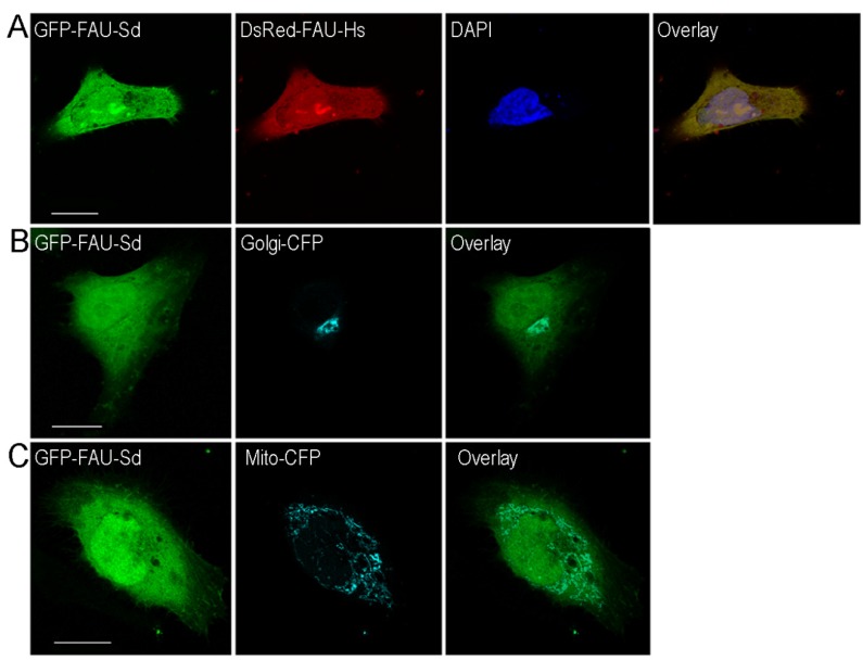
Figure 4.
(A) Subcellular localization of sponge and human FAU. HeLa cells transiently transfected with sponge (Sd) pEGFP-FAU (green fluorescence), human (Hs) pDsRed-FAU (red fluorescence); (B) pECFP-Golgi (cyan) and (C) pECFP-mitochondria (cyan). The overlay (yellow) shows colocalization of the human and sponge homologs in panel A. Scale bar = 10 μm.

