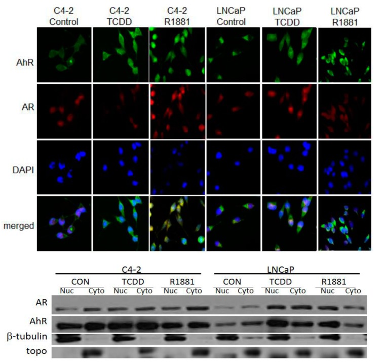
Figure 2.
IHC staining of LNCaP and C4-2 prostate cancer cells. Cells were exposed to 1 μM TCDD or 10 nM R1881 for 24 h. AhR was visualized by staining with FITC-conjugated goat anti-rabbit antibody and AR with Rhodamine-conjugated rabbit anti-mouse antibody. The nuclei were counter-stained with DAPI fluorescence dye. Images from FITC, Rhodamine and DAPI-fluorescence channels were merged (A). Western blotting of nuclear (nuc) and cytoplasmic (cyto) fractions. Cells were exposed to 1 μM TCDD or 10 nM R1881 for 24 h and isolated fractions were transferred to a PVDF membrane which was probed with AhR and AR antibodies. β-tubulin was used as a cytoplasmic loading control and topoisomerase (topo) was used as a loading control for nuclear fractions (B).

