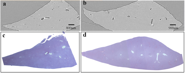
Figure 2. Histological sections and CT images of the normal and mild liver samples.

The PCI-CT images in (a) normal and (b) mild liver fibrosis samples are presented, and the corresponding histological sections are shown in (c) and (d). The magnification of histological section is 12.5×.
