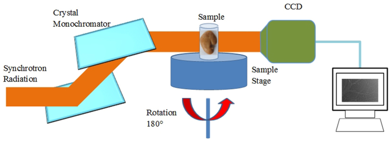
Figure 6. Schematic diagram of the PCI experimental setup at BL13W1 in SSRF.

A monochromatized synchrotron radiation x-ray is projected on a sample mounted on a rotating sample stage, and the transmitted beam is recorded by an image detector after the x-ray propagates a proper distance from the object. For tomographic scans, the sample can be rotated within 180° to produce the projection images at different views.
