
Figure 4. AOSLO image shows patchy loss of cone outer segments in left temporal retina, far from retina corresponding to the enlarged blind spot.
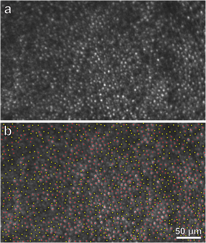
(a) Zone centered at 6.5° temporal to the fovea (red box, Fig. 3), at a mirrored location to the box in Fig. 5a, showing many absent cone outer segments. (b) Identification of 549 missing (yellow dots) and 650 intact (red dots) outer segments in (a). The region measures 0.10 mm2, yielding a total density of 11,990 cones/mm2, with 6,500 intact cones/mm2 and 5490 missing cones/mm2. The density in the normal right retina at the same eccentricity is 12,290 visible cones/mm2 (Fig. 5e,f). Despite loss of nearly half the cones, retinal sensitivity was depressed by only a half log unit (3–6 dB) (Fig. S3).