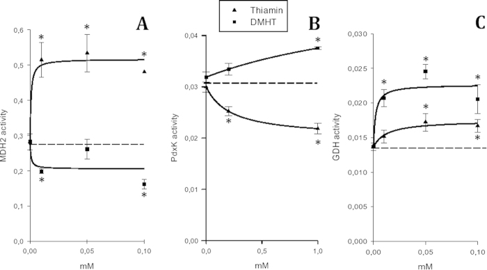
Figure 2. Influence of thiamin and DMHT on activities of the enzymes abundant in the thiamin and thiazolium proteomes.
(A)—mitochondrial malate dehydrogenase (MDH2) assay in Ringer-bicarbonate buffer, pH 7.4, at 0.01 mM oxaloacetate and 0.02 mM NADH; (B)—human recombinant pyridoxal kinase (PdxK) assay in 75 mM NaBES, pH 7.3, at 0.125 mM pyridoxal and 0.1 mM ATP; (C)—glutamate dehydrogenase (GDH) assay in Ringer-bicarbonate buffer, pH 7.4, at 0.1 mM 2-oxoglutarate and 0.02 mM NADH. Activities are expressed in micromoles of substrate transformed per min per mg of protein. Each data point represents the average ± SEM from at least triplicate assays. When error bars are not visible, they are within the symbol size. The experimental curves were approximated by hyperbolic functions using SigmaPlot 12.0. Statistical significance (p ≤ 0.05, t-test) of the differences compared to the control values is marked by asterisks.

