Figure 2.

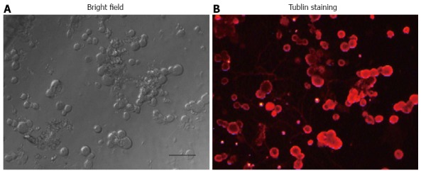
Bright-field of acutely dissociated dorsal root ganglion cells (A), and image of the same dorsal root ganglion cells (B) in (A) stained with β-tubulin (red). Bar = 50 μm.

Bright-field of acutely dissociated dorsal root ganglion cells (A), and image of the same dorsal root ganglion cells (B) in (A) stained with β-tubulin (red). Bar = 50 μm.