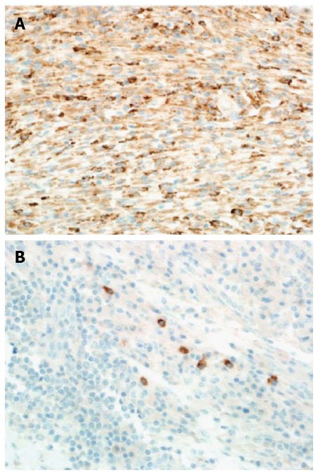
Figure 7.

Immunohistochemical analysis of the focal hepatic lesion. A: All histiocytes in the lesion stained diffusely positive for CD-68 (HE stain, magnification power: × 40). B: A few plasma cells in the lesion stained focally positive for IgG4 (IgG4 stain, magnification power: × 40).
