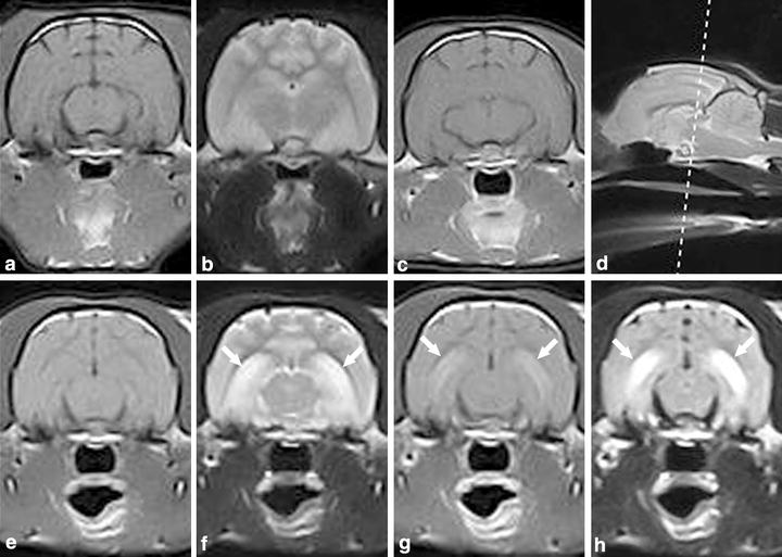
Figure 1.

Case 1. a–c First MRI examination. A-T1W transversal, B-T2W transversal, C-T1W with contrast transversal magnetic resonance images. Normal appearance of the brain. Case 1. e–h Second MRI examination of the brain 32 months after epilepsy onset. E-T1W transversal, F-T2W transversal, G-T1W with contrast transversal, H-FLAIR transversal magnetic resonance images. Arrows indicate bilateral hyperintensity in the hippocampus in T2W + FLAIR and contrast enhancement in T1W + C. Magnetic resonance imaging findings consists of moderate to marked bilateral symmetric hyperintense lesions in T2W and FLAIR. Moderate to marked contrast enhancement is seen bilaterally in the hippocampus. d D-T2W sagittal magnetic resonance image with reference line located in rostral midbrain indicating where the transversal slices were obtained.
