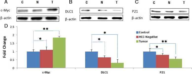
Fig. 2.

Oncoproteins: Western blots of the control, HCC-negative and liver tumor tissues (as is shown in Fig. 1) were probed with antibodies against c-Myc, DLC-1 or p21 proteins (panels a, b and b). Panel (d) represents quantitative analysis (based on the loading controls) of liver tissues from uninfected control, HCC negative and HCC positive mice (as in Fig. 1)
