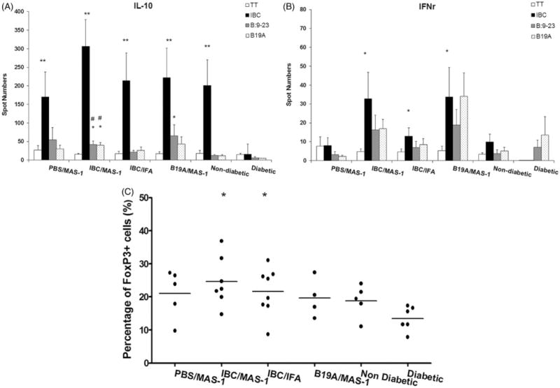
Figure 3.

Antigen-specific IL-10- (A) and IFN-γ- (B) producing splenocytes of 52-week old diabetes-free NOD mice immunized with PBS/MAS-1, IBC/MAS-1, IBC/IFA or B19A/MAS-1 and re-stimulated ex vivo with PBS (open bar) insulin peptides IBC (black bar), B:9-23 (gray bar) and B19A (dot bar) measured by ELISPOT assay. Non-diabetic NOD mice at 25–30 weeks and new onset DM (within one week post diagnosis) were also analyzed for comparison. (C) The percentage of FoxP3+ population of CD4+ splenocytes from diabetes protected mice at 52 weeks. Each dot in (C) represents each surviving mouse. Statistical significance of <0.001 is denoted by **, * and #. *p≤0.005 and **p≤0.001 compared with new onset DM; #p≤0.05 and ##p≤0.001 compared with non-diabetic NOD mice.
