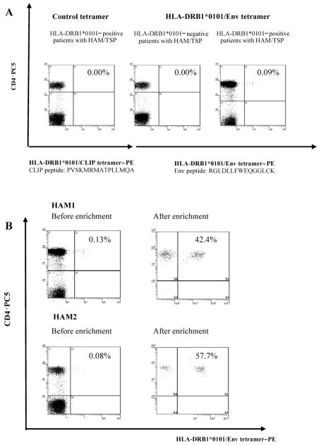
Figure 1.
Direct ex vivo detection and enrichment of human T lymphotropic virus type 1 (HTLV-1) Env gp21–specific CD4+ T cells by the DRB1*0101/Env380-394 (DR1/Env) tetramer. A, No detectable staining of tetramer+ peripheral blood mononuclear cells in DRB1*0101-positive patients with HTLV-1–associated myelopathy/tropical spastic paraparesis (HAM/TSP) by the control HLA-DRB1*0101/CLIP tetramer (left panel) or in DRB1*0101-negative patients with HAM/TSP by the DR1/Env tetramer (center panel). In contrast, clear and distinct staining was observed in DRB1*0101-positive patients with HAM/TSP by the DR1/Env tetramer (right panel). All DR1/Env tetramer+ cells were CD4+. The percentages of cells in each quadrant are given in the upper right corner. B, Ex vivo major histocompatibility complex class II tetramer staining of HTLV-1 Env380-394 –specific CD4+ T cells (left panels). DR1/Env tetramer+ T cells were positively selected from negatively selected CD4+ T cells by use of anti-phycoerythrin (PE) MACS beads and a magnetic separation column (right panels). The purity was checked by flow cytometry with anti-CD4 monoclonal antibody staining. PC5, cyanine 5–succinimidylester.

