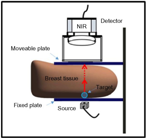
Figure 3.

Setup for breast imaging studies consisting of the breast tissue placed in between two transparent plates.
Notes: A detector is placed above the top plate, the source is placed beneath the bottom plate, and a target is placed beneath the breast tissue and above the bottom plate.
Abbreviation: NIR, near-infrared.
