Figure 2.
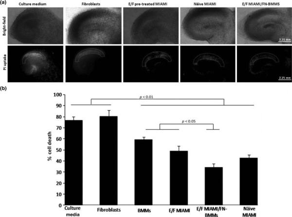
A) Representative images of hippocampal slice cultures. Bright-field and propidium iodide fluorescence images taken 24h after the lethal ischemic (40 min of OGD) insult of culture medium, fibroblasts, EGF/bFGF-pretreated MIAMI cells, naïve MIAMI and EGF/bFGF pre-treated MIAMI/FN-BMMs, respectively. B) Propidium fluorescence values measured in the CA1 pyramidal cells in rat organotypic slices one day after ischemia. BMMs, E/F pre-treated MIAMI cells, E/F pre-treated MIAMI cells/FN-BMMs and näive MIAMI cells were neuroprotective as compared with culture medium and human fibroblasts-injected group (p<0.01). E/F pre-treated MIAMI cells/FN-BMMs were more neuroprotective as compared to BMMs and E/F pre-treated MIAMI (p<0.05). % Cell death as defined in the methods, reflects the ratio of propidium iodide staining 24h after lethal ischemia (OGD) and propidium iodide staining 24 h after 100 μm/L NMDA treatment (total cell death).

