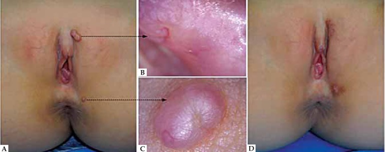
FIGURE 1.
A. A 9-year-old girl developed a pink raised nodule of 0.5 X 1.1cm in diameter and a reddish 0.4 X 0.3cm papule around the vulva and anus respectively. B. Dermoscopic view of the nodule showed its smooth, reddish and piny surface with small shallow depressions. C. Under dermoscopy, a depression in the margin of the pink papule, on which a crown vessel was apparent. D. The lesions were completely removed by an excisional biopsy

