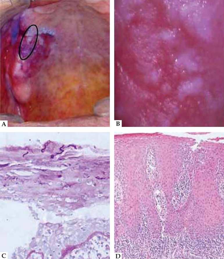
FIGURE 1.
A. Clinical case of OLP associated with candidiasis. Presence of a 2.0x2.0cm red area with white plaques, irregular contour and indefinite boundaries, located in the upper alveolar ridge and right hard palate; B. Clinical case of OLP associated with candidiasis. Image of the lesion captured by a intraoral camera at 28x magnification; C. Clinical case of OLP associated with candidiasis. PAS staining. Presence of Candida spp. hyphae; D. Clinical case of OLP associated with candidiasis. HE staining at 100x magnification: presence of severe exocytosis, degeneration of the basal layer, dysplastic changes such as irregular epithelial stratification, mitosis, mild hyperchromatism. These changes affected the basal and parabasal layers, and were consistent with moderate dysplasia and intense mononuclear infl ammatory infiltrate.

