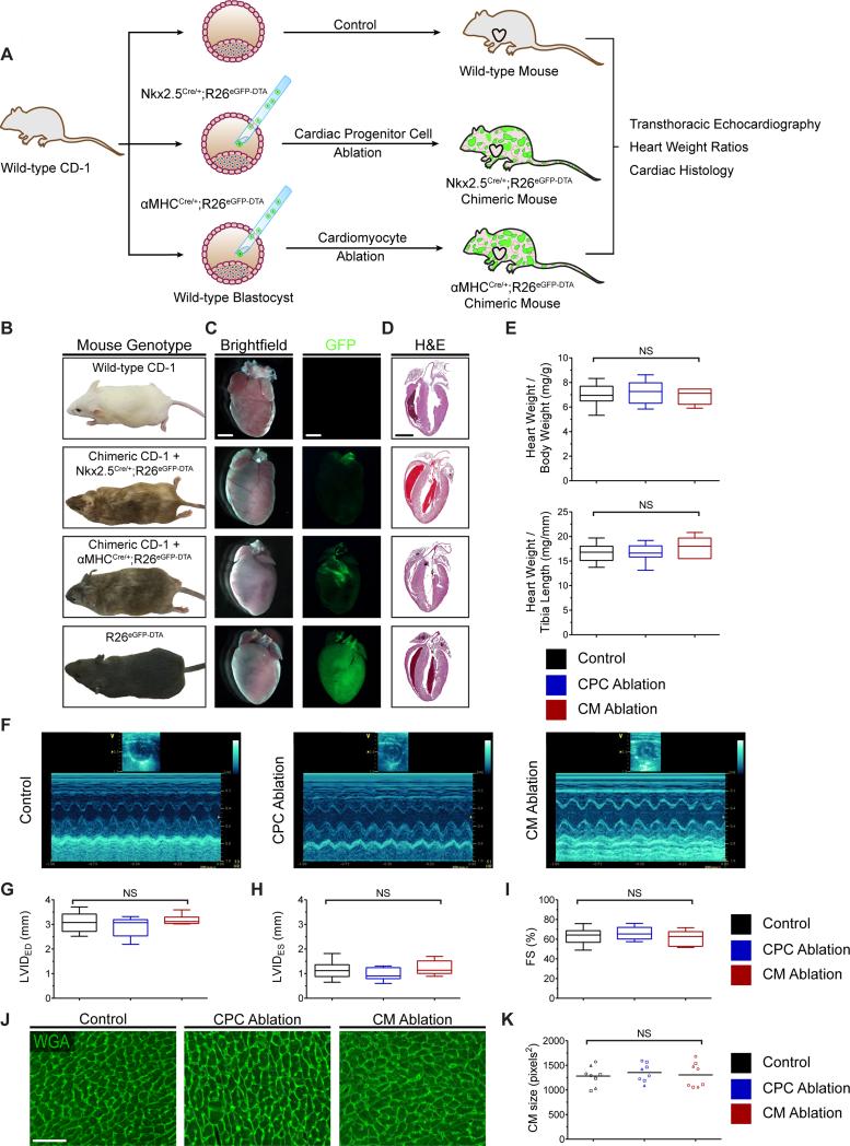
Figure 5.
CPC and CM-ablated embryos can survive into adulthood with no overt cardiac defect. (A) Strategy used for generation and analysis of 4-5 week-old mice undergoing CPC or CM ablation. (B) Representative images of control and ablated chimeric adult mice. (C) Brightfield and GFP fluorescence images of the dissected whole hearts. GFP signal is nearly absent in the hearts that have undergone ablation. (D) H&E staining of representative heart sections from control and ablated hearts. For (E-K): experimental variables are represented as follows: [group: # of mice examined, mean percent ablation ± SD]. For (E-I): results are displayed using box-and-whisker plots demonstrating median value and interquartile range. Whiskers span the minimum and maximum range of values. (E) Heart weight to body weight ratio and heart weight to tibia length ratio in wild-type and ablated hearts [control: n=28; CPC ablation: n=11, 39 ± 9%; CM ablation: n=6, 42 ± 11%] (F-I) Representative M-mode echocardiography images (F) and left ventricular dimensions (G and H) and function (I) quantified by fractional shortening [control: n=16; CPC ablation: n=9, 40 ± 10%; CM ablation: n=7, 41 ± 10%]. (J and K) Wheat germ agglutinin (WGA) staining of control and ablated hearts (J) and dot plot (K) displaying mean CM size. Each symbol in the dot plot represents the mean CM size in an individual heart section; a minimum of three 32× fields per section were counted; each symbol shape represents an independent mouse; a horizontal bar indicates the mean value for each group. [control: n=3; CPC ablation: n=3, 51 ± 6%; CM ablation: n=3, 51 ± 7%]. FS, fractional shortening; LVIDED, left ventricular internal diameter at end-diastole; LVIDES, left ventricular internal diameter at endsystole; NS, non-significant. Scale bars: 2 mm for (C and D); 50 μm for (J).

