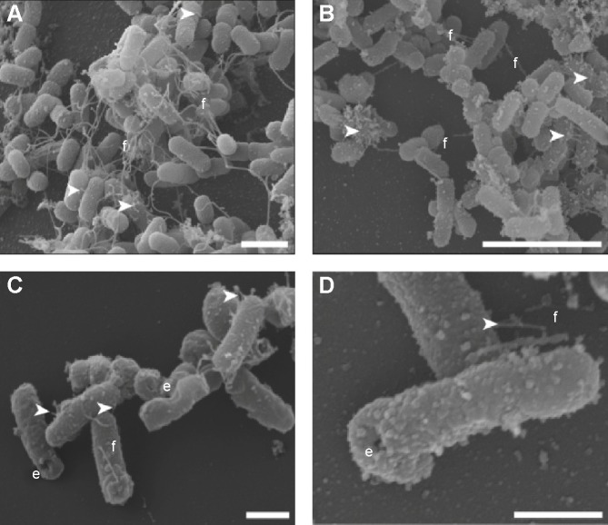
Figure 1.
Scanning electron microscopic images, showing both EcN and EcN BGs expressing foreign chlamydial antigen.
Notes: (A) N-PmpC-EcN during exponential growth showing abundant flagellae and fimbriae. (B, C, and D) N-PmpC-EcN BGs after E-lysis showing retention of flagellae (f) and fimbriae (white arrowheads). The E-lysis pore is visible (e) on some of the EcN BGs. Scale bars: (A) 2 µm; (B) 5 µm; (C) 1 µm; and (D) 500 nm.
Abbreviations: BGs, bacterial ghosts; EcN, Escherichia coli strain Nissle 1917; N-PmpC, N-terminal part of polymorphic membrane protein C.

