Figure 2.
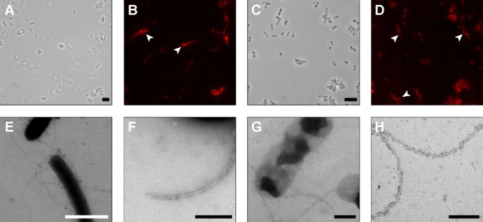
Flagella of both EcN and EcN BGs were identified using immunohistochemistry and immunoelectron microscopy.
Notes: (A) EcN (pBGKB-MOMP, pGLysivb) bacteria, transmitted light; (B) flagella of EcN (pBGKB-MOMP, pGLysivb) are localized and labeled with Alexa Fluor 546 (white arrowheads); (C) MOMP-EcN (pBGKB-MOMP, pGLysivb) BGs, transmitted light; (D) flagella of MOMP-EcN (pBGKB-MOMP, pGLysivb) BGs are localized and labeled with Alexa Fluor 546 (white arrowheads); (E) TEM image of EcN (pBGKB-MOMP, pGLysivb) bacteria, showing negatively contrasted flagella labeled with 25 nm colloidal gold; (F) high-magnification TEM image of EcN (pBGKB-MOMP, pGLysivb) bacteria, showing negatively contrasted flagella labeled with 6 nm colloidal gold; (G) lower magnification and (H) high-magnification TEM image of MOMP-EcN (pBGKB-MOMP, pGLysivb) BGs showing negatively contrasted flagella labeled with 12 nm colloidal gold. Scale bars: (A–D) 5 µm; (E) 2 µm; and (F–H) 500 nm.
Abbreviations: BGs, bacterial ghosts; EcN, Escherichia coli strain Nissle 1917; MOMP, major outer membrane protein; TEM, transmission electron microscope.

