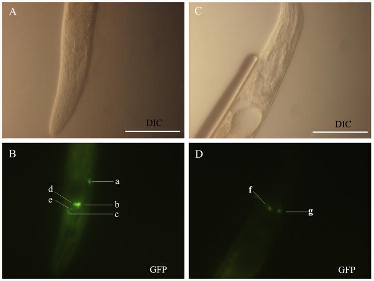
Fig. 6.

Representative expression profiles displayed in Caenorhabditis elegans using two GFP constructs, pL-CG2 and pL-HG2 (cf. Fig. 1). (A and B) Differential interference contrast (DIC) and fluorescence images of a N2 (wild type) L3 using the construct Ce-daf-2 p∷gfp (pL-CG2), respectively. GFP reporter expression was present in the head neuron (a), amphidial neurons including ASH (b), ADF (d) and AWA (e), and nerve ring (c). (C and D) DIC and fluorescence images showing the expression of construct Haemonchus contortus Hc-daf-2 p∷gfp (pL-HG2) in L3 stage of a N2 C. elegans. GFP reporter expression was present in amphidial neuron AWA (f and g). Scale bars = 50 μm.
