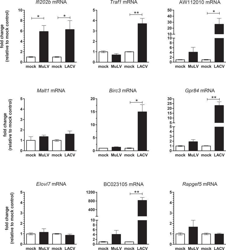
Fig 6. Real-time PCR analysis of mRNA expression of selected genes in brain tissue of mice with viral encephalitis.
Brain tissue from mice with signs of neurological disease following infection with MuLV or LACV was processed for RNA. Age-matched and strain-matched controls for each virus infection were processed at the same time as viral infection and are shown as controls for the respective viruses. RNA was then analyzed for expression of mRNAs of genes identified as being induced following TLR activation of microglia and/or astrocytes. Data are the mean +/- SEM of 3–6 mice per group and are shown as the fold change relative to the average mock sample for each group. Statistical analysis was completed by unpaired t test between the virus-infected brain tissue and the appropriate mock-infected control. P<0.05, ** P<0.01, *** P<0.001.

