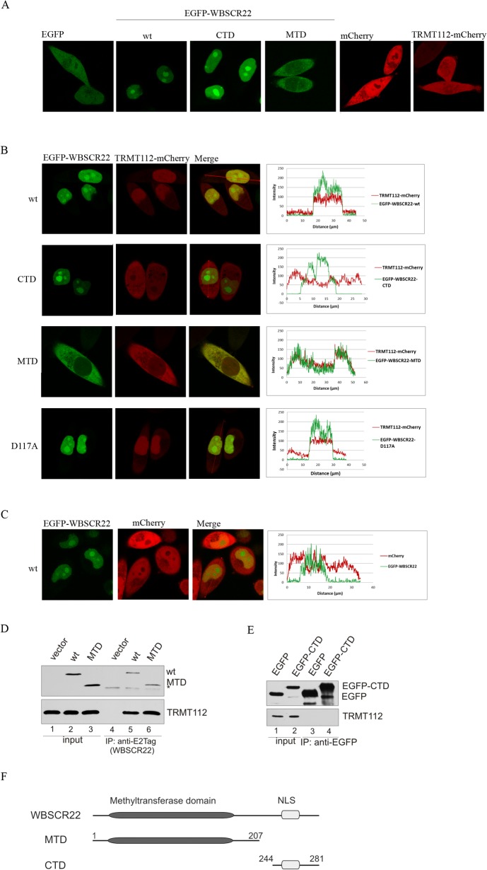
Fig 3. Localization of TRMT112 is determined by the WBSCR22 protein.
(A) Live cell confocal images of EGFP, EGFP-WBSCR22, EGFP-WBSCR22-CTD, EGFP-WBSCR22-MTD, mCherry and TRMT112-mCherry proteins in HeLa cells. (B) Live cell confocal images and fluorescence intensity profile graphs of TRMT112-mCherry protein co-expressed with EGFP-WBSCR22, EGFP-WBSCR22-CTD, EGFP-WBSCR22-MTD and EGFP-WBSCR22-D117A, and (C) mCherry protein co-expressed with EGFP-WBSCR22. Fluorescence signal was visualized using confocal laser scanning microscope LSM710 (Zeiss). Images were obtained with 63x lens and analyzed by ZEN2011 software. (D) Co-immunoprecipitation of WBSCR22 and TRMT112 proteins. COS-7 cells were transfected with plasmids encoding for WBSCR22 and its mutant MTD. 24 hours later co-immunoprecipitation was performed using antibody against E2Tag. Immunoblotting was performed with antibodies against E2Tag (HRP-conjugate) and TRMT112. The non-specific signal is shown by asterisk. (E) Co-immunoprecipitation of EGFP-tagged WBSCR22-CTD protein. Immunoprecipitation was performed with magnetic beads that were covalently coupled with EGFP binding protein and analyzed by immunoblotting with antibodies against EGFP and TRMT112. (F) A schematic representation of WBSCR22 proteins.

