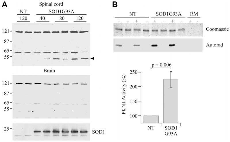
Fig. 5.
PKN1 is cleaved and activated in SOD1G93A transgenic mice. (A) Immunoblots to show PKN1 in spinal cord (upper) and brain (lower) from SOD1G93A and non-transgenic littermate (NT) mice at ages 40, 80 and 120 days. Full-length PKN1 (120 kDa) and an approx. 65 kDa species are seen in all samples. An additional approx. 55 kDa migrating species (arrowhead) is seen in spinal cord but not brains of SOD1G93A mice at 80 and 120 days; this species is not detected in non-transgenic littermates. The samples were also probed with an antibody to SOD1 to confirm the genotype (bottom panel). Two mice at each age are shown but an additional two mice were analysed with highly similar results. (B) shows in vitro kinase assays for PKN1 activity in spinal cords of SOD1G93A and non-transgenic littermate (NT) mice. Both the Coomassie stained gel showing substrate and corresponding autoradiograph are shown along with reaction mix controls with no substrate (RM). + and − refer to presence or absence of the PKN1 antibody in the immunoprecipitations to isolate PKN1. PKN1 activity is elevated approximately 2.25-fold in SOD1G93A mice (n = 8, t-test).

