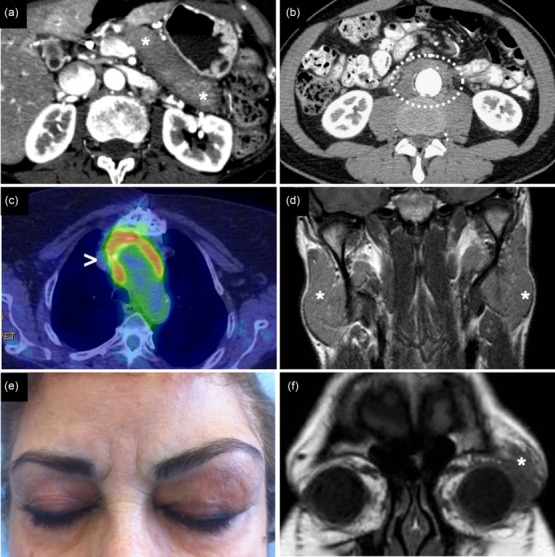
Figure 1.

Clinical and radiological presentation of IgG4-related disease (IgG4-RD). (a) IgG4-related autoimmune pancreatitis: computed tomography scan showing a ‘sausage-like’-shaped pancreas with a surrounding rim of hypodense tissue (asterisks). (b) Retroperitoneal fibrosis with periaortic involvement (circle). (c) Inflammatory aneurism of the thoracic aorta showing 18fluoro-deoxyglucose uptake on positron emission tomography (arrowhead). (d) Magnetic resonance showing bilateral parotid enlargement due to IgG4-RD (asterisks). (e,f) Clinical and radiological appearance of IgG4-related orbital pseudotumour (asterisk).
