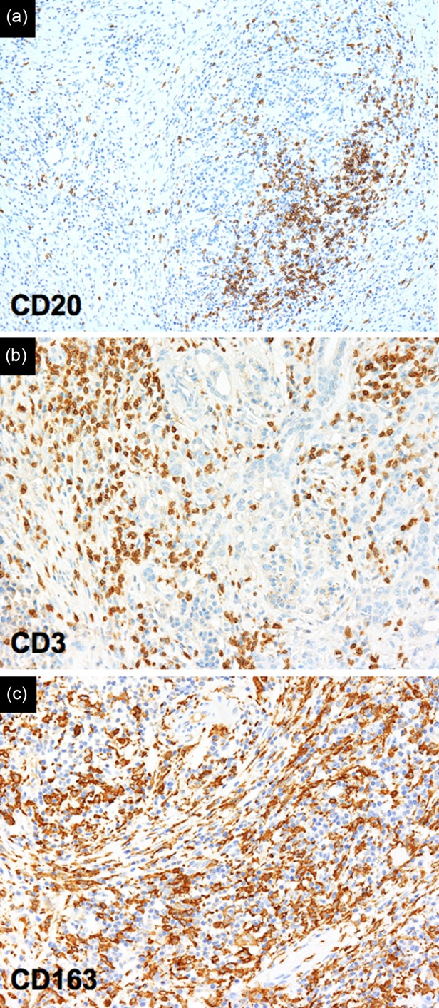
Figure 3.

Inflammatory infiltrate in IgG4-related disease. Immunohistochemistry reveals CD20+ B lymphocytes organized in follicular structures (a), CD3+ T lymphocytes (b), and CD163+ M2 macrophages (c) spread throughout the fibrotic tissue (magnification ×100).
