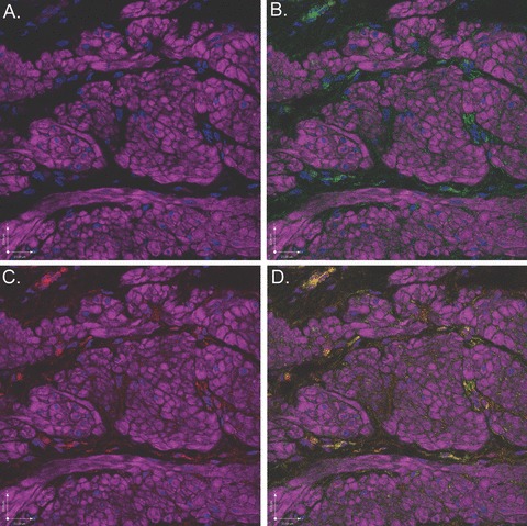
Figure 11.

Scanning confocal microscopic images of rabbit bladder sections processed using FIHC reveal COX-1 expression in c-kit-positive ICCs surrounding DSM bundles. In all panels, purple staining (phalloidin) demonstrates DSM bundles and blue staining (DAPI) demonstrates nuclei. (A) Image in which staining for c-kit and COX-1 is withheld as a control revealing DSM bundles. (B) Identical image in which c-kit (green) identifies ICCs surrounding DSM bundles. (C) Identical image in which COX-1 (red) identifies ICCs surrounding DSM bundles. D. Overlay image of (B) and (C) in which yellow staining reveals co-localization of c-kit and COX-1 in ICCs. This is a representative picture from n= 3 bladders.
