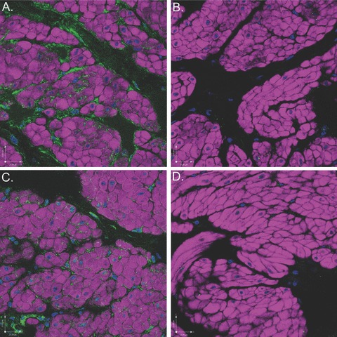
Figure 12.

Scanning confocal microscopic images of rabbit bladder sections processed using FIHC indicate near abolishment of COX-2 and COX-1 staining in the presence of specific BP. All images are dual-stained with phalloidin (purple) to demonstrate DSM morphology and DAPI (blue) to demonstrate nuclear morphology. (A) COX-1 expression (green) on ICCs surrounding DSM bundles. (B) COX-1 expression is nearly abolished in the presence of COX-1 BP. (C) COX-2 expression (green) on ICCs surround DSM bundles. (D) COX-2 expression is nearly abolished in the presence of COX-2 BP.
