
Figure 13.
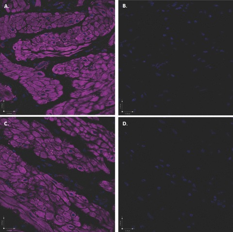
Scanning confocal microscopic images of rabbit bladder sections processed using FIHC indicate an absence of non-specific labelling with secondary antibodies. All images were labelled with phalloidin (purple) and DAPI (blue) and were exposed to the four excitation wavelengths described in the methods and corresponding to the colours blue (405 nm), green (488 nm), red (568 nm) and purple (647 nm). (A) Addition of secondary antibody Alexa Fluor 488 (without primary antibody) reveals no evidence of non-specific green staining. (B) In the identical image, digital subtraction of the purple colour does not uncover any subtle non-specific green staining in the region of the DSM bundles. (C) In an additional section labelled with phalloidin and DAPI, addition of the secondary antibody Alexa Fluor 568 (without primary antibody), reveals no evidence of non-specific red staining. (D) In the identical image, digital subtraction of the purple colour does not uncover any subtle non-specific red staining.